Yong Fang1,
Liang Lv2,
Yong-Jian Chai3,
Fan Yu1 ![]()
For correspondence:- Fan Yu Email: yufany3@gmail.com Tel:+8653189029000
Received: 20 September 2016 Accepted: 25 November 2016 Published: 20 December 2016
Citation: Fang Y, Lv L, Chai Y, Yu F. Neuroprotective effect of whole-plant extract of Torilis leptophylla in isoflurane-treated rats. Trop J Pharm Res 2016; 15(12):2571-2578 doi: 10.4314/tjpr.v15i12.6
© 2016 The authors.
This is an Open Access article that uses a funding model which does not charge readers or their institutions for access and distributed under the terms of the Creative Commons Attribution License (http://creativecommons.org/licenses/by/4.0) and the Budapest Open Access Initiative (http://www.budapestopenaccessinitiative.org/read), which permit unrestricted use, distribution, and reproduction in any medium, provided the original work is properly credited..
Purpose: To investigate the neuroprotective effect of Torilis leptophylla (TL) extract on animals exposed to isoflurane-induced anesthesia.
Methods: General anesthesia was induced in diabetic rats by administration of 2 % sevoflurane (v/v) in 100 % oxygen. The animals from the treatment group were orally administered TL extract. Behavioral parameters, the explicit objection recognition index, and performance in water maze experiments were observed. Additionally, the neuroprotection provided by the herbal extract was measured in terms of the reduction in oxidative stress in isolated animal brain tissue.
Results: The study showed approximately 1.5-fold improvement in object recognition index of TL-extract-treated anesthetized rats compared with the control group of anesthetized animals. Additionally, the escape latency values of the anesthetized rats in Morris water maze experiments were reduced following TL extract treatment at all time points throughout the 5-day study protocol. Finally, biomarkers of oxidative stress in the brain indicate that the levels of superoxide dismutase and catalase enzymes were improved by approximately 1.43- and 1.36-fold in TL-extract-treated rats compared with those in the control group.
Conclusion: The results of the present study show that cognitive dysfunction and brain injury associated with enhanced oxidative stress can be reversed by the antioxidant properties of the TL extract.
Introduction
General anesthesia in patients of different age groups, ranging from infants to geriatrics, with varying pathological conditions and mechanical trauma has been a common medical practice for several decades [1,2]. Among the various anesthetic drugs, isoflurane has been the most widely utilized inhalational anesthetic agent.
Isoflurane is a transparent, stable substance with a feebly pungent musty odor. It is supplied as a 99.9 % pure liquid with no pharmaceutical additives. Isoflurane is known to produce its pharmacological action by agonistic binding to gamma aminobutyric acid (GABA), glutamate, and glycine-type receptors. In addition, it antagonizes N-methyl-D-aspartate (NMDA) receptor activity. Isoflurane is one of the most popular inhalational general anesthetics (Forane®) used to induce anesthesia, particularly in pediatric patients, because of its quick onset of action and rapid recovery. Moreover, isoflurane is less irritating to the airways than other anesthetic agents. However, some recent studies have depicted setbacks associated with the use of isoflurane in experimental subjects, particularly regarding younger age groups. The detrimental effects of isoflurane-induced anesthesia comprise neurodegeneration and cognitive impairment resulting mainly from increased oxidative stress and inflammatory response mediators.
This prompted us to investigate the prophylactic potential of Torilis leptophylla (TL) extract on young rats exposed to isoflurane-induced anesthesia. TL is a plant known as bristle fruit hedge parsley, which is mainly found in several regions of Asia, Europe, and North Africa [3]. Historically, TL has been known for its anti-inflammatory and antimicrobial properties. It is widely used in traditional medicine for the treatment of gastrointestinal disorders and as a general tonic. Recent studies have suggested the presence of multiple antioxidant components in TL [3,4]. Despite the holistic therapeutic applications of the TL plant, its potential seems largely unexplored in modern medicine.
Therefore, the current study aimed to investigate the prophylactic properties of a whole-plant extract of TL in reducing the extent of cognitive impairment and brain injury in neonatal subjects. Wistar rats were used as animal subjects for our studies. Various evaluations were performed using behavioral and biochemical parameters.
Methods
Materials
TL plant was obtained in August 2014 from Shanghai Botanical Gardens (Shanghai, China). Authentication of the plant material was performed by Dr Feng Shucheng, Professor of Plant Taxonomy, Shanghai Botanical Gardens. A voucher specimen (accession no. YF-014408) was deposited at the Herbarium of Shanghai Botanical Garden for future reference. Isoflurane was procured from a local pharmacy store in Shandong, China while streptozotocin was obtained from Sigma-Aldrich (Shanghai, China). The other reagents and chemicals used were of analytical grade.
Preparation of extract
All of the plants (5 kg) were dried under shade to obtain 1 kg of dry sample, which was milled in a 7.5-kg ball-mill (JHMA®; Weifang Jinghua Ltd., Shadong, China) to obtain a coarse powder. Thereafter, 250 g of the coarse powder was extracted twice with 1,500 mL of 95 % ethanol at room temperature for 24 h. The solvent was concentrated in a rotary evaporator (Rotavapor® R215; Buchi Shanghai Ltd., Shanghai, China) under reduced pressure at 40 °C to yield the TL extract.
Animals
The experimental protocol was conducted after approval of the Institutional Animal Ethics Committee of Jinan Maternity and Child Care Hospital (approval ref no. 201208342) and was performed in compliance with Directive 2010/63/EU on careful handling of animals for scientific purposes [5]. Male neonatal Wistar rats (postnatal day 14) were randomly assigned to different study groups and were housed at 22 °C in polypropylene rat cages (9 animals per cage) [6]. The animals were kept in a light-controlled environment with a 12-h light-dark cycle and were allowed continuous access to a rat pellet diet.
Treatment with the TL extract
Before starting the anesthesia, the TL extract was administered to the rats in the treatment group for 3 days. The extract was provided as twice daily oral doses separated by 12 h (5 mL/kg body weight) using an oral cannula feeder. The animals receiving the TL extract were referred to as the TL-extract-treated group.
Isoflurane-induced anesthesia
The rats (n = 18) were placed in plexiglass sealed chambers maintained at a temperature of 37 ± 1°C. The chambers comprised a provision for 1.5 % isoflurane in a gaseous mixture of 35 % O2/65 % N2 (1 L/min). The rats were exposed to isoflurane for 5 h. Following the anesthesia phase, the animals were transferred to another glass chamber with a provision of oxygen to attain recovery for 1 h. The TL extract-treated animals exposed to isoflurane anesthesia were termed the TL-ISF-treated group, whereas naïve rats receiving anesthesia were called as the ISF-treated group. The rats in the ISF-control group were only administered pure oxygen (without anesthesia) and were termed the SVF-control group. Twenty-four hours after inducing anesthesia, a subgroup of animals (n = 6) from each treatment group was sacrificed to collect blood samples for biochemical analysis. The remaining animals were subjected to behavioral determinations after 32 days.
Object recognition test
A plastic tank (60 cm × 60 cm × 25 cm) was used in the studies, and the animals were placed in the tanks for at least 10 sessions of 15 min for 2 days before initiating the studies. The object recognition experiments were performed in two phases. First, the rats were individually placed in the arena, wherein two identical objects were already placed. The time taken to explore the objects was recorded. The arena was thoroughly cleaned in-between such sessions. Object exploration refers to the incidence of sniffing (at less than 2 cm) and prolonged rearing on the object (> 5 s). The second phase of the study comprised replacing of 1 of the 2 identical objects with a different object (different color, odor, and texture). The rats were again placed in the arena, and the exploration time was recorded. The test parameter was the recognition cognitive index [7], calculated as in Eq 1.
Recognition index = [Td/(Td+Tf)] x 100 …… (1)
where Td and Tf represent the time taken to explore different objects and the time taken to explore familiar objects, respectively.
Morris water maze test
The Morris water maze test was performed using a conventional Morris maze (Shanghai Jiliang Ltd., Shanghai, China). The swim pattern, path length, and time needed to reach the platform were recorded by a video camera and analyzed by integrated computer software. Prior to commencing the studies, the rats were trained for 1 day to swim and rest on a visible platform destination. Similarly, the rats were subsequently trained to swim and rest on a submerged (hidden) platform for the next 2 days. After completion of the swim training, the Morris water maze test was performed for 5 consecutive days. The animals were transferred from four different locations in the maze and were allowed to search for the platform for 2 min; if the animal failed to find the platform, they were helped to the platform and kept there undisturbed for another 30 s. The parameters observed were the time spent in the quadrants (Qtime) and the escape latency, which refers to the time taken by the animal to find the platform.
Collection of brain tissue samples
The rats were sacrificed by decapitation. Tissue from the hippocampus was homogenized in radioimmuno-precipitation assay (RIPA) buffer using a tissue homogenizer (PRO250® tissue homogenizer; Pro-Scientific Inc., Oxford, CT, USA), and the homogenates were centrifuged at 4000 × g for 15 min. The supernatants were isolated and utilized for several estimations [8,9].
Determination of oxidative stress biomarkers
The protein content was quantified in the tissue homogenates using Gornall’s biuret technique, with bovine serum albumin (BSA) as the reference protein [10]. Briefly, each tissue homogenate sample (0.1 mL) was mixed with sodium chloride (2.9 mL) and working biuret reagent (3.0 mL). This mixture was allowed to rest at room temperature for 30 min, and its absorbance was recorded at 540 nm in a double-beam UV spectrophotometer (Shimadzu UV-200; Shimadzu, Osaka, Japan). Thereafter, oxidative stress was estimated in terms of superoxide dismutase (SOD), lipid peroxidation (expressed as malondialdehyde, MDA), and catalase (CAT).
Superoxide dismutase (SOD)
Freshly prepared aqueous solutions of nitroblue-tetrazolium chloride (1.0 mL, 7.8 %) and hydroxylamine hydrochloride (1.0 mL, 13.9 %) were added to 0.1 mL of the tissue homogenate. Thereafter, the change in absorbance was recorded at 560 nm over 120-s intervals [11].
Malondialdehyde (MDA)
Acetic acid (1.5 mL, 20 %), aqueous sodium lauryl sulfate (0.2 mL, 8.1 %), aqueous thiobarbituric acid (1.5 mL, 0.8 %), and triple-distilled water (0.6 mL) were added to the tissue homogenate (0.2 mL). The mixture was exposed to heat (70 °C) and then allowed to cool gradually. Afterward, 1 mL of triple-distilled water and an n-butanol:pyridine mixture (15:1 v/v; 5 mL) was added and agitated vigorously until the organic layer separated. The organic layer was isolated using a phase separation funnel, and its absorbance was measured at 532 nm [12].
Catalase (CAT) activity
The catalase activity in the tissue homogenates was measured using Thiele’s method [13]. The disappearance of hydrogen peroxide (H2O2) is an indication of the consumption of CAT. Hydrogen peroxide (30 mmol/L) was mixed along with the tissue homogenate, and changes in the absorbance values were monitored at 240 nm for 120-s intervals. CAT level was derived from Eq 2.
CAT (K/min) = 2.3/∆t × logA1/A2 ……..………. (2)
where A1 and A2 represent the initial and final absorbance values, and the time interval ∆t is 60 s.
Statistical analysis
All of the data were analyzed using one-way analysis of variance (ANOVA) and expressed as mean ± standard deviation (SD); p < 0.05 was considered to be statistically significant. Statistical analysis was performed using Graph-Pad Prism® 4.0 (Graph-Pad Software Inc., San Diego, CA, USA).
Results
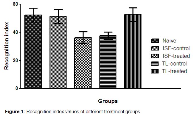
Identical recognition index values were observed for naïve rats and ISF-control rats (55.25 ± 4.05 and 51.29 ± 5.09, respectively) (), which reflected the physiological memory function of healthy rats. Significantly lower (p < 0.05) recognition index values of ISF-treated rats (36.21 ± 4.18) indicated that exposure to anesthesia caused significant cognitive impairment in neonatal rats. The animals exposed to blank extract solvents (TL-control) showed no improvement in cognitive impairment, whereas those treated with TL extract showed tremendous improvement in cognitive function. The animals that received the phytochemical extract exhibited full replenished object recognition ability, as indicated by the high recognition index value (52.63 ± 3.88).
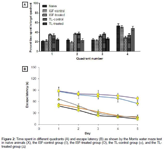
The results of the water maze study are shown in A and B. Post-training, the naïve rats and those from the ISF-control group spent the maximum Qtime in the fourth quadrant, which was used as the target quadrant throughout the studies. Additionally, the minimum values for the escape latency was on Day 1 in naïve and STN-control animals, values that were further reduced with time. Post-anesthesia, the animals from the ISF-treated group exhibited noticeable cognitive impairment as evidenced by the significant (p < 0.05) reduction in Qtime in the target quadrant. Additionally, a reduced Qtime in the target quadrant was observed in animals in the TL-control group. On the other hand, the animals treated with TL extract showed a markedly improved Qtime in the target quadrant, indicating cognitive replenishment in the young rats.
The escape latency exhibited by SVF-treated animals was also significantly higher (p < 0.05) than that of naïve and ISF-control animals. After ISF-induced anesthesia, the cognitive loss in neonatal rats (ISF-treated group) seemed to be more prominent than that in all other groups as the lowest value for Qtime in the fourth quadrant was observed. Furthermore, the escape latency values were significantly higher (p < 0.05) during all of the study days.
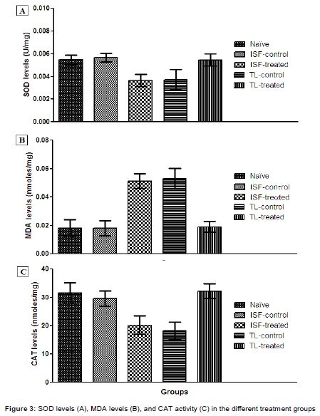
The levels of oxidative stress biomarkers in the various groups are shown in . It was observed that rats treated with anesthesia (ISF-treated) showed a significant increase (p < 0.05) in the MDA levels compared with naïve and ISF-control animals. On the other hand, the levels of SOD and CAT were significantly reduced (p < 0.05) after the administration of anesthesia compared with those in the naïve and ISF-control animals.
Discussion
Isoflurane exposure has long been known to produce neurodegenerative effects rapidly in neonatal experimental rats [7,14]. These effects are especially critical because isoflurane is the most widely used general anesthetic in neonatal and young patients.
Isoflurane has been used as a general anesthetic worldwide for several years. Recent studies have depicted the neurodegenerative effects of anesthesia induced using isoflurane. Bonin and Orser [14] reported severe tau-phosphorylation of hippocampal tissue after 1 h of isoflurane administration in mice, leading to significant memory loss. On the other hand, Yang et al [13] demonstrated short-term memory loss associated with isoflurane-induced anesthesia in rats [14], and Leopke et al [15] emphasized isoflurane-related cognitive impairment in neonatal animals.
Additionally, surgical procedures in diabetic subjects are crucial. Very recently, the antioxidant and neuroprotective functions of TL extract have been reported [3,4]. The antioxidant properties of the plant could provide an excellent prophylactic effect in neonatal patients, which are required to be anesthetized. Therefore, the current study was aimed to evaluate the potential of TL extract in preventing cognitive and neurophysiological dysfunction in neonatal Wistar rats as animal subjects.
It was observed in the water maze test as well as in the object recognition test that isoflurane-induced anesthesia in healthy animals (ISF-treated group) as well as in placebo-treated animals (TL-control group) produced significant memory loss with respect to naïve animals.
This indicated that no pharmacological activity was shown by the placebo-treated animals. As previously stated, isoflurane has been widely accepted to increase the symptoms of anesthesia-related cognitive dysfunction, which can be attributed to several factors, such as isoflurane-induced neuronal damage, the over-expression of mediators of inflammation, and increased oxidative stress in the hippocampal region of the brain [15–17]. The observations of the current study indicate that the isoflurane-induced neurodegeneration and cognitive loss in anesthetized neonatal animals was considerably reduced upon treatment with TL extract.
To determine the injury resulting from oxidative stress due to isoflurane administration, the effect of TL extract on biomarkers of oxidative stress was studied. SOD is a defensive enzyme, which causes dismutation of toxic superoxide free radicals into oxygen and hydrogen peroxide. Similarly, CAT is also a defensive enzyme, which catalyzes toxic hydrogen peroxide molecules into nontoxic water and oxygen. Decreases in SOD and CAT levels are an indicator of increased oxidative stress or brain cell injury. On the other hand, the formation of MDA indicates a detrimental effect of reactive oxygen species on brain lipoproteins, and the end products are commonly termed lipoxidation end products. Therefore, high MDA levels indicate severe brain cell injury in isoflurane-treated rats. The anesthetized animals showed certain signs of increased oxidative stress and brain injury. However, when the animals were provided prophylactic treatment in the form of TL extract, the oxidative stress was noticeably less, indicating a reduction in neurodegeneration.
Conclusion
General anesthesia induced by isoflurane can produce significant impairment of the cognitive behavior of neonatal subjects, likely attributable to the enhancement of oxidative stress in the hippocampal region of the brain. Oral treatment with TL extract, however, provides an efficient prophylaxis in neonatal rats exposed to isoflurane anesthesia. This study suggests that TL is potentially a reasonable phyto-constituent to counter isoflurane-related brain injury in higher mammals.
Declarations
Acknowledgement
References
Archives


News Updates